



Birthmarks removal in Kyiv
Why it is necessary to examine
Removal methods
Why exactly Esteva Clinic
Current prices and promotions
- Examination of neoplasms by experienced dermatologists
- Effective removal methods: radio wave, cryodestruction, CO2 laser
- Fast healing without scar
What are birthmarks
A mole is a skin growth containing melanocytes (specialised skin cells) and the pigment melanin. Their colour can range from light brown to black. Moles are also known as nevi.
Every mole goes through a specific development cycle. Often, a nevus first appears as a small, flat spot, but gradually becomes raised. The height of such a growth depends on the location of the melanocytes: if they are in the upper layers of the skin, the mole will be flat; if they are located deeper, it will rise above the surface of the skin. This structure is merely a characteristic; it has no negative significance. However, the structure and appearance of the growth should be monitored more closely.
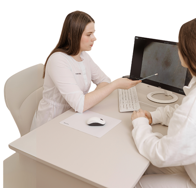
What you need to know about moles and their treatment
Moles are rare on babies. Usually, the main number of them appears in adolescence and adulthood. There are several reasons for the formation of new birthmarks on humans:
- heredity;
- the sun and UV radiation (excessive exposure to direct sunlight or in a solarium leads to excessive production of melanin, which causes birthmarks). Most often, new birthmarks appear precisely because of the influence of sunlight on the skin;
- hormonal changes (both in teenagers and in adults);
- viruses and injuries.
Why histology is important
Histological examination is the study of a tissue sample at the cellular level. The results of the histological examination confirm or deny the previous diagnosis, determine the tactics of treatment of the disease. This procedure is used to diagnose cancer and other diseases.
This type of examination is indicated for all patients who consult a dermatologist for the removal of a suspicious nevus or neoplasm that is often injured and bleeds. Sending mole tissues for histological examination is mandatory.
Dermatoscopy - what it is and why it is performed
Dermatoscopy is the most common diagnostic method, which is performed using a dermatoscope medical device. The procedure allows you to examine birthmarks, to reveal the degree of their threat to human health. The dermatoscope magnifies the image tenfold, which allows you to carefully study the skin formation, determine the size, exact shape, color, and also consider the structure. Based on the results of dermatoscopy, the doctor makes a conclusion as to whether it is possible to leave the mole or whether it is better to remove it.
The procedure is absolutely painless and harmless. It should be performed annually by every person who has moles. This diagnosis is especially indicated for owners of a large number of moles that differ in color and size. Regular diagnostics help to identify changes in the structure of moles and, if necessary, to solve the problem in the early stages.
Why moles need to be examined and sometimes removed
A mole is, by its very nature, a benign growth. It develops from immature or altered cells of the epidermis. In most cases, a mole poses no threat to life or health and affects only the aesthetic perception of a person’s appearance. However, in some cases, these growths may eventually increase in size, penetrate deeper, and change in structure; in such cases, the nevus may pose a threat not only to health but also to life.
To determine how dangerous a mole is, there is the ABCDE test:
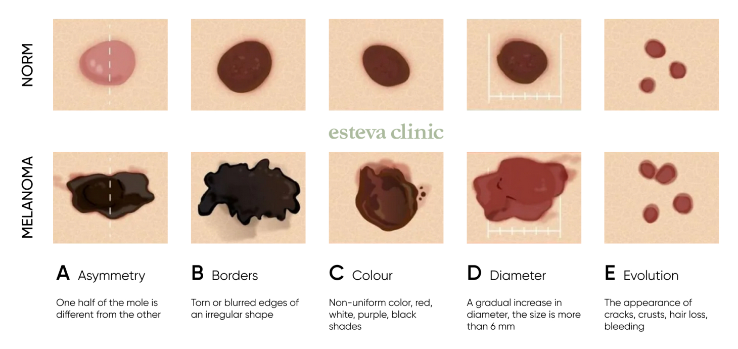
- A is “asymmetry”. The birthmark changes shape and becomes asymmetrical.
- B is “borders”. In a good-quality education, the contour should be smooth, not torn.
- C is “colour”. A safe formation has a brown color, a uniform tone over the entire surface. If the nevus becomes red, white, dark purple, black or initially colored in several colors at once, it should be studied in more detail.
- D is “diameter”. Moles should have certain, fixed sizes. If it gradually increases in diameter, this may indicate pathological processes.
- E is “evolution”. A change in the shape, structure or size of the skin is meant.
Regular monitoring of your moles, accurate diagnosis and the removal of unwanted growths can help to protect your health and save your life. You should consult a dermatologist as soon as you notice the first changes in the appearance of a mole. The doctor will carry out a visual examination, perform a dermatoscopy and, based on the results of the examination, provide recommendations on how to proceed.
Which moles should be removed?
All moles can be divided into two main categories: benign and malignant.
Benign moles generally do not need to be removed unless there are specific medical reasons for doing so. However, benign moles can become malignant if certain safety precautions are not followed (in particular, exposure to strong ultraviolet and radiofrequency radiation).
Malignant moles pose a certain risk and require close monitoring. They are often removed because, when it comes to moles, the sooner the problem is addressed, the more likely a positive outcome is and the lower the risk of developing a highly dangerous condition – melanoma.
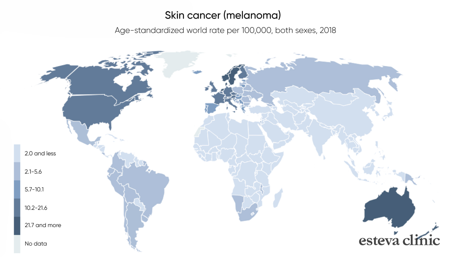
A mole should be removed if:
- it has changed shape, size or colour;
- there is inflammation or swelling around it;
- the structure has become compromised, with flaking or scabbing;
- the texture of the mole has changed – it has become either too firm or, conversely, much softer;
- it has a large diameter from the outset – over 6 mm;
- the mole causes psychological or physical discomfort;
- the mole is located in an area of the body where it is constantly rubbed by clothing.
How to treat moles properly
The simplest way to get rid of a mole is to have it removed. This is also the most effective method. What’s more, it’s quick: the procedure itself takes just a few seconds. It is essential to consult a dermatologist beforehand: the specialist will need to assess the nature of the growth and select the most appropriate method of removal. In some cases, a histological examination is carried out prior to removal to obtain further information. The dermatologist will also provide recommendations on skin care during the recovery period.
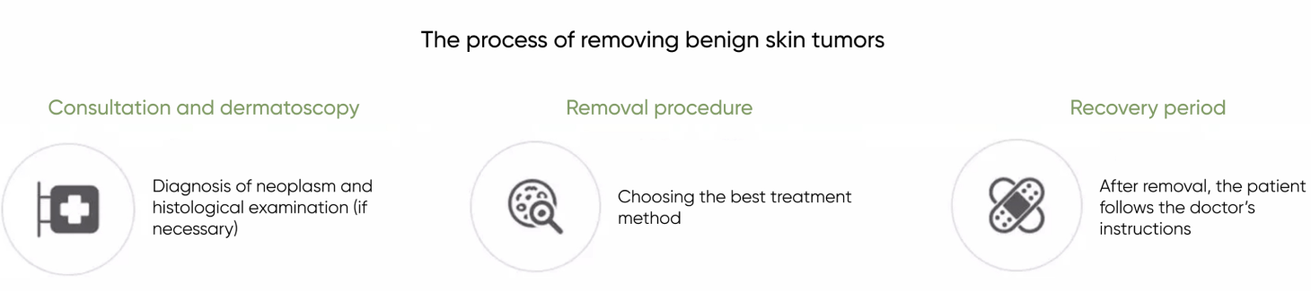
How is the removal carried out?
Regardless of the method chosen (radiofrequency or CO₂ laser), it will definitely be:
- fast;
- painlessly;
- safety;
Understanding how the procedure to remove benign growths will be carried out helps patients to remain calm and keep a positive outlook.
We have tried to answer the most frequently asked questions we receive on this topic. We have compiled all the answers into a video, which we invite you to watch.
The active part of the process takes less than a minute!
How to remove a mole or nevus
At Esteva Clinic, we offer the most effective methods for removing skin lesions. Currently, these include removal using radio waves, liquid nitrogen (cryodestruction) or a CO₂ laser.
Radio wave method
With this method, radio waves are directed at the affected area, destroying the cells of the mole. As a result of this treatment, the mole shrinks in size until it disappears completely. The radio wave method of removing growths can be used not only on the skin, but also on more delicate areas – such as mucous membranes.
20 minutes before the procedure, a local anaesthetic is applied to the affected area – this helps to prevent any discomfort.
The radiofrequency treatment then begins. When removing growths on the arms or legs, the doctor may, at their discretion, recommend an additional procedure known as curettage (scraping of the tissue). This ensures that the nevus has been completely removed.
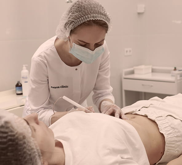
The skin recovers quickly—within a week after the procedure.
Advantages of the radiofrequency method:
- the epidermis surrounding the skin remains intact;
- local anaesthesia completely eliminates any pain.
The radiofrequency method is suitable for removing moles in the following areas:
- face;
- décolletage, chest, underarms,
- arms (including the palms).
Laser method
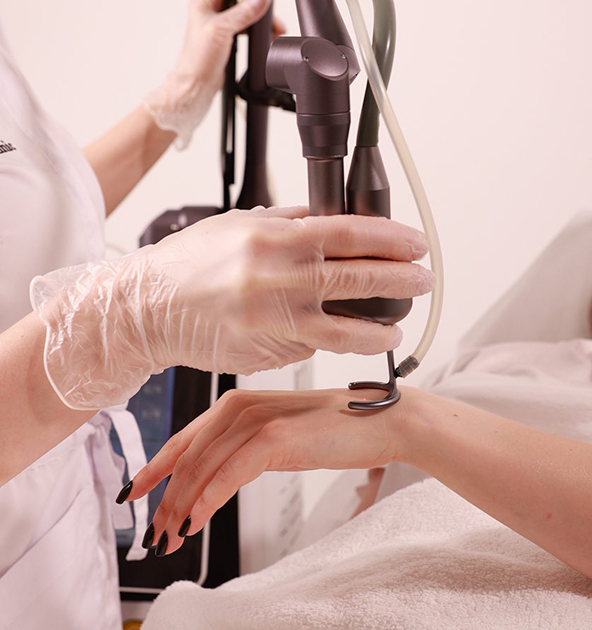
One of the most popular technologies today. The removal principle is similar to the radiofrequency method, with the sole difference being that a laser beam is used here. The healthy skin surrounding the growth is not affected, and no scars remain after the procedure.
Laser mole removal is particularly common on difficult-to-reach areas:
- on the fingers and toes, in the immediate vicinity of the nail plates;
- on the soles of the feet, particularly on the plantar surface.
Laser and radiofrequency treatments are two of the ‘gold standard’ methods for removing not only moles but also other skin growths, such as papillomas, haemangiomas and warts.
Mole removal – before and after photos
Frequently asked questions about mole removal
Is a histological examination or dermatoscopy required before removing moles?
A histological examination is mandatory in the following cases:
- the presence of traumatised skin lesions;
- before removing moles, nevi and polyps
So, in short, yes, moles are removed using dermatoscopy and histology
More details
Surgical excision of a mole using a scalpel is considered the most reliable option in situations where there is a greater than 20% likelihood of the growth becoming malignant. This method is also advisable for large growths (over 0.5 cm), those that have grown deeply into the tissue, or if the nevus is regularly injured or subjected to friction, for example, from clothing.
More details
Mole removal at Esteva Clinic will not significantly disrupt your normal routine. You can resume most of your daily activities immediately after the procedure.
The mole is removed completely. However, if the underlying cause of the mole’s appearance remains in the body, it is quite possible that a new mole (nevus) will appear nearby or elsewhere on the body.
Both methods of mole removal are effective. A dermatologist will advise which is more suitable in a particular case following a dermatoscopic examination.
The benefits of mole and nevus removal at Esteva Clinic
- We are experts in our field – our team consists exclusively of experienced dermatologists.
- Esteva Clinic offers three main methods for removing benign skin growths: radiofrequency treatment, cryotherapy and CO₂ laser treatment. These are quick, painless and bloodless removal methods.
- The clinic offers digital dermatoscopy and histology.
- Esteva Clinic is an official partner of the Skin Checker social initiative (by La Roche-Posay), which aims to combat malignant skin tumours.
How much does it cost to have a mole removed?
Видалення родимок та невусів
| Назва процедури | Ціна, грн |
|---|---|
| Консультація дерматолога обов’язкова | 990 |
| Контроль після видалення з 30 по 45 день | Безкоштовно |
| Родимки, травмовані новоутворення, невус, поліпи - видалення з гістологією новоутворення до 1 см | 2300 |
| Родимки, травмовані новоутворення, невус, поліпи - видалення з гістологією новоутворення до 2 см або на повіці | 3300 |
| Родимки, травмовані новоутворення, невус, поліпи - видалення з гістологією новоутворень до 1 см до 3 шт | 5900 |